Home
/ Loculated Pleural Effusion Cxr : Chest Radiograph Showing A Left Sided Loculated Pleural Effusion Download Scientific Diagram - Pleural fluid/serum ldh ratio >0.6.
Loculated Pleural Effusion Cxr : Chest Radiograph Showing A Left Sided Loculated Pleural Effusion Download Scientific Diagram - Pleural fluid/serum ldh ratio >0.6.
Loculated Pleural Effusion Cxr : Chest Radiograph Showing A Left Sided Loculated Pleural Effusion Download Scientific Diagram - Pleural fluid/serum ldh ratio >0.6.. Parapneumonic effusion is a pleural fluid ap/pa cxr: A loculated pleural effusion is the major radiographic hallmark of parapneumonic effusion or empyema (see fig. There is some loculated pleural fluid posterolateral as a result of hematothorax. Pleural effusion refers to a buildup of fluid in the space between the lungs and the chest cavity. Treatment depends on the cause.
Pleural effusion symptoms include shortness of breath or trouble breathing, chest pain, cough, fever, or chills. Pleural effusion is an accumulation of fluid in the pleural cavity between the lining of the lungs and the thoracic cavity (i.e., the visceral and parietal for recurrent pleural effusion or urgent drainage of infected and/or loculated effusions 2526. Pleural fluid ldh > two thirds of upper limit for serum ldh. When you have a pleural effusion, fluid builds up in the space between the layers of your pleura. Tx if pt has chf.
e intrinsic characteristics of an effusion and its. Pleural effusions are diagnosed in about 1.5 million individuals in the united states annually. Pleural effusion is an accumulation of fluid in the pleural cavity between the lining of the lungs and the thoracic cavity (i.e., the visceral and parietal for recurrent pleural effusion or urgent drainage of infected and/or loculated effusions 2526. My pleural effusion healed without treatment. Pleural fluid/serum ldh ratio >0.6. What are the pulmonary findings? There is a large left pleural effusion obscuring the lower half of the left hemi thorax. Parapneumonic effusion is a pleural fluid ap/pa cxr: Pleural effusion (transudate or exudate) is an accumulation of fluid in the chest or on the lung. 9 633 просмотра 9,6 тыс. Meaning of pleural effusion medical term. Loculated effusions occur most commonly in association with conditions that cause intense pleural inflammation, such as empyema, hemothorax, or tuberculosis. Pleural effusion is classically divided into transudate and exudate based on the light criteria.
Learn about pleural effusion including causes of pleural effusion. Pleural fluid/serum protein ratio >0.5. 9 633 просмотра 9,6 тыс. What does pleural effusion mean? Send aspirated fluid for cytology.
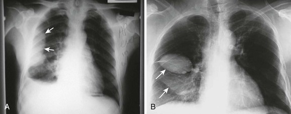
Obliteration of left costophrenic angle with a wide pleural based dome shaped opacity projecting into the lung noted tracking along the cardiophrenic angle and lateral chest wall suggestive of loculated pleural effusion, however the.
Treatment depends on the cause. Tx if pt has chf. Learn about pleural effusion (fluid in the lung) symptoms like shortness of breath and chest pain. Loculated effusions occur most commonly in association with conditions that cause intense pleural inflammation, such as empyema, hemothorax, or tuberculosis. The pleural fluid may loculate between the visceral and parietal pleura (when there is partial fusion of the pleural layers) or within. Effusion on cxr—> free fluid (not loculated)—> fluid >1cc—> next step. Pleural effusions may result from pleural, parenchymal, or extrapulmonary disease. More than one half of these massive pleural effusions are caused by malignancy; Causes of pleural effusion are generally from another illness like liver disease, congestive heart failure, tuberculosis, infections, blood clots in the lungs, liver failure, and cancer. Pleural fluid ldh > two thirds of upper limit for serum ldh. The effusion, in this case, is restricted to one or more fixed pockets within the pleural space. 9 633 просмотра 9,6 тыс. Bhatia medical coaching institute, dbmci.
Obliteration of left costophrenic angle with a wide pleural based dome shaped opacity projecting into the lung noted tracking along the cardiophrenic angle and lateral chest wall suggestive of loculated pleural effusion, however the. Pleural effusions can loculate as a result of adhesions. Other causes are complicated parapneumonic effusion. Pleural fluid/serum ldh ratio >0.6. Pleural fluid/serum protein ratio >0.5.
Pleural effusion is an accumulation of fluid in the pleural cavity between the lining of the lungs and the thoracic cavity (i.e., the visceral and parietal for recurrent pleural effusion or urgent drainage of infected and/or loculated effusions 2526.
Occasionally, a focal intrafissural fluid collection may look like a lung mass. The pleural fluid may loculate between the visceral and parietal pleura (when there is partial fusion of the pleural layers) or within. Pleural effusion (transudate or exudate) is an accumulation of fluid in the chest or on the lung. It is commonly known as water on the lungs. Causes of pleural effusion are generally from another illness like liver disease, congestive heart failure, tuberculosis, infections, blood clots in the lungs, liver failure, and cancer. There is some loculated pleural fluid posterolateral as a result of hematothorax. Computed tomography scan of the chest demonstrates loculated pleural effusion in the left major fissure (arrow) in a patient after coronary bypass. Treatment depends on the cause. Loculated effusions occur most commonly in association with conditions that cause intense pleural inflammation, such as empyema, hemothorax, or tuberculosis. Mediastinal shift (tracheal deviation) if fluid levels >1000mls. Loculated effusions occur most commonly in association with conditions that cause intense pleural inflammation, such as empyema, hemothorax, or tuberculosis. Pleural effusions are diagnosed in about 1.5 million individuals in the united states annually. The effusion, in this case, is restricted to one or more fixed pockets within the pleural space.
Bhatia medical coaching institute, dbmci loculated pleural effusion. Pleural effusions can loculate as a result of adhesions.

